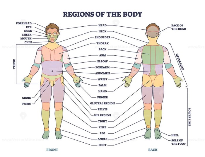
Correctly label the following regions of the external anatomy. – Correctly labeling the following regions of the external anatomy is a fundamental aspect of anatomical study and medical practice. Standardized terminology and precise labeling are crucial for effective communication among healthcare professionals and accurate diagnosis and treatment of patients. This guide provides a comprehensive overview of the external anatomy, including the head and neck, thorax, abdomen, limbs, and back, empowering readers with the knowledge to confidently identify and label these regions.
Throughout history, anatomical labeling conventions have evolved to ensure clarity and consistency in describing the human body. By adhering to these conventions, we can avoid confusion and facilitate precise communication in clinical settings.
External Anatomy Overview: Correctly Label The Following Regions Of The External Anatomy.

Correctly labeling external anatomical regions is crucial for effective communication and understanding in the medical field. Standardized terminology ensures that healthcare professionals can accurately describe and locate anatomical structures, facilitating accurate diagnosis, treatment, and research.
The history of anatomical labeling conventions dates back to ancient times, with notable contributions from early anatomists such as Galen and Leonardo da Vinci. Over time, these conventions have been refined and expanded to encompass the comprehensive labeling system used today.
Head and Neck Regions, Correctly label the following regions of the external anatomy.
The head and neck region encompasses several distinct anatomical areas. The face is divided into the forehead, cheeks, nose, mouth, and chin. The eyes, ears, and scalp are also located in this region.
The neck is divided into the anterior and posterior triangles. The anterior triangle contains the larynx, trachea, esophagus, and major blood vessels. The posterior triangle contains the muscles and vertebrae of the neck.
Thorax and Abdomen Regions
The thorax, or chest, is enclosed by the ribs and sternum. It contains the heart, lungs, and major blood vessels. The diaphragm separates the thorax from the abdomen.
The abdomen is divided into four quadrants by two imaginary lines: the midclavicular line and the transpyloric plane. It contains the stomach, intestines, liver, gallbladder, and pancreas.
Limbs and Appendages
The upper limbs consist of the shoulder, arm, forearm, wrist, and hand. The major bones of the upper limbs are the humerus, radius, and ulna.
The lower limbs consist of the hip, thigh, leg, ankle, and foot. The major bones of the lower limbs are the femur, tibia, and fibula.
Back and Spine Regions
The back is divided into the neck, shoulders, thoracic region, lumbar region, and sacral region. The major muscles of the back include the trapezius, latissimus dorsi, and erector spinae.
The spine, or vertebral column, consists of 33 vertebrae stacked one upon another. The spine provides support and protection for the spinal cord and nerves.
Surface Anatomy Techniques
Surface anatomy techniques, such as palpation and auscultation, allow healthcare professionals to examine and assess anatomical structures through the skin.
Palpation involves using the hands to feel for anatomical landmarks, such as bones, muscles, and organs. Auscultation involves listening to sounds produced by anatomical structures, such as the heart and lungs.
Clinical Applications
Accurate anatomical labeling is essential for medical diagnosis and treatment. It enables healthcare professionals to precisely locate and describe anatomical structures, ensuring that interventions are targeted and effective.
In surgical procedures, anatomical labeling guides the surgeon’s incisions and manipulations. In imaging techniques, such as X-rays and CT scans, anatomical labeling helps radiologists interpret the images and identify any abnormalities.
Popular Questions
Why is it important to correctly label the external anatomy?
Correctly labeling the external anatomy is crucial for effective communication among healthcare professionals, accurate diagnosis and treatment of patients, and precise documentation in medical records.
What are the key anatomical landmarks of the head and neck?
Key anatomical landmarks of the head and neck include the eyes, nose, mouth, ears, trachea, esophagus, and major blood vessels.
How can I improve my ability to label the external anatomy?
Practice regularly using anatomical models, diagrams, and cadavers. Attend anatomy classes and workshops, and consult reputable anatomy textbooks and online resources.

